|

|
|
Jung Y. Huang received his Ph.D in 1988 from Applied Physics of Cornell University. Currently he is a principal investigator and professor of the T.K.B. Research Center for Photonics, Chiao Tung University, Taiwan. Research Opportunities Available for Postdocs and Ph.D Students: We are developing real-time single-molecule light sheet microscopy for exploring the dynamical signaling networks in live cells. We are endeavoring to decipher the molecular events of those challenging problems, such as how extracellular signals trigger the formation of dynamic structures of receptors at the plasma membranes and how the signals are relayed to downstream molecules and affect relevant gene expressions. If you are interested in this project, please hit Two scientific revolutions will celebrate their 100th and 200th anniversaries in 2015, respectively. Learn what they are. |
|

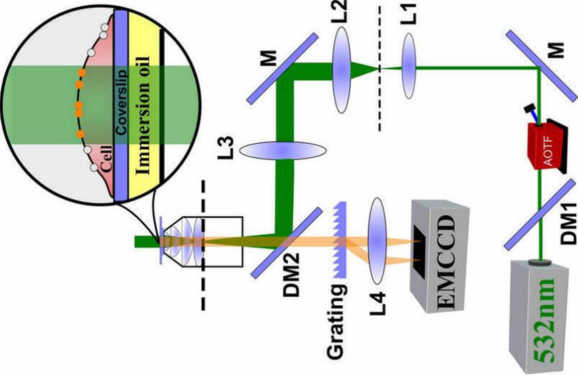
Research Objectives(1) Imaging Subcellular Dynamic Processes in Live Cells with Time-lapse Light Sheet MicroscopyIn recent years, live cell optical imaging has become instrumental for studying cellular function and processes. To explore those important dynamic processes, we developed a time-lapsed light-sheet microscope. The apparatus is equiped with a fast image-taking software based on mixed CPU/GPU implementation to allow for real-time single-molecule 3D localization. The setup depicted in Fig. 1(a) can produce a diffraction-limited ligh sheet in z direction. The profile of the light sheet was presented in Fig. 1(c), revealing a beam waist of 2.9 μm and a Rayleigh length of 41 μm. In the illumination arm, a 2D scanner combined with an electric tunable lens (ETL) (not shown) can move the light sheet by 34 μm along the y and z directions, and 420 μm along the x direction at the sample position. The positioning accuracy is about 0.5 μm. 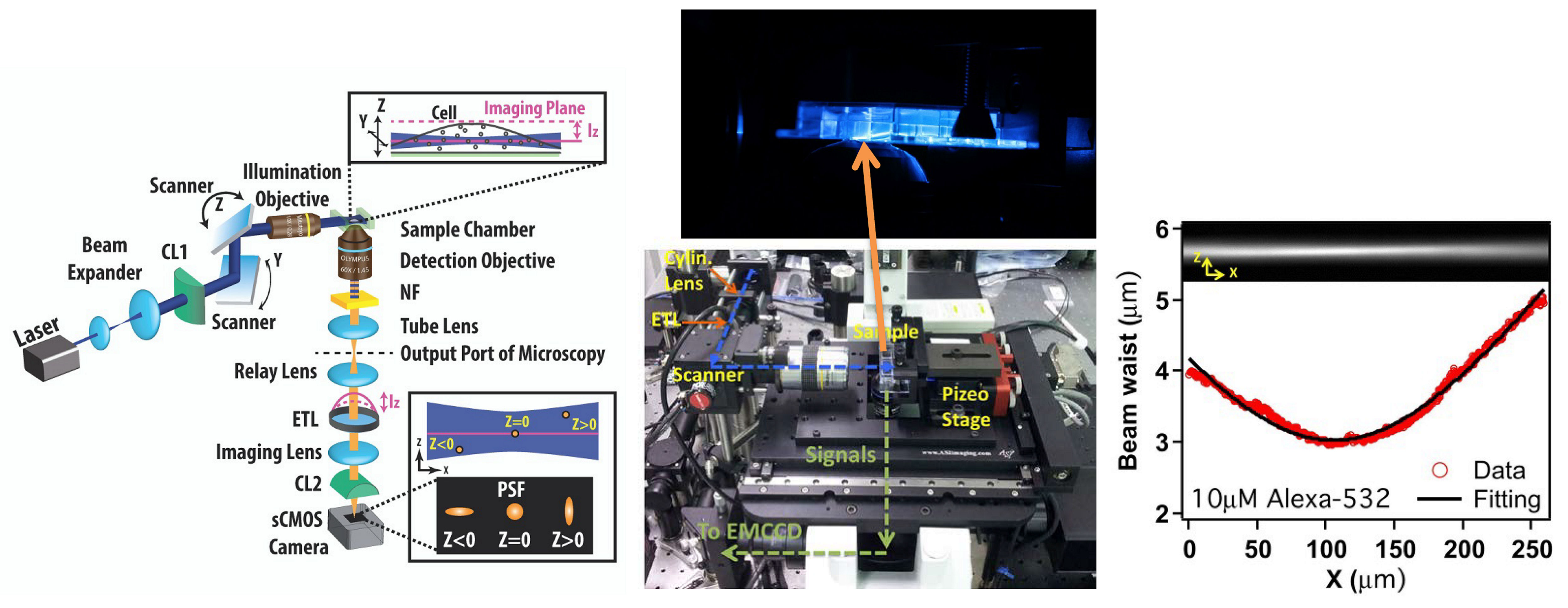
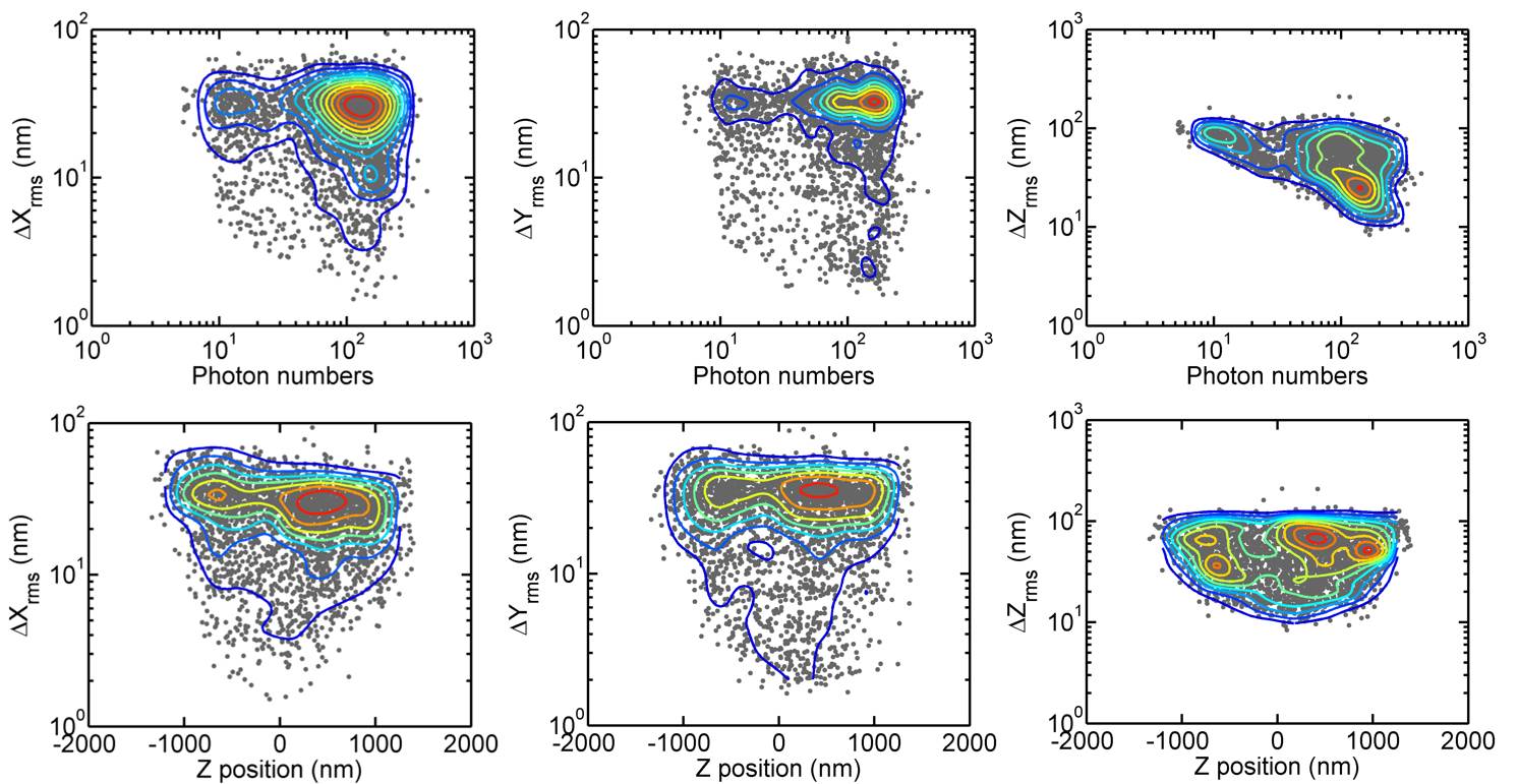
Fig. 1: (Left) Schematic of the time-lapsed light-sheet microscope, (Middle) Image of the setup around the sample cell and the neighboring 2D scanner, illumination and detection objective lenses, (Right) the light sheet revealed by the fluorescence from a 10-uM Alexa-532 solution. For a given position of the light sheet, we can adjust the ETL of the imaging arm to scan the focal plane to yield z-resolved images of the light-sheet illuminated sample. We also used the astigmatism introduced by CL2 to encode the depth information of a fluorophore into the elliptically shaped PSFs as shown in Fig. 2. In this way, the axial position of a flurophore within a distance of 1000 nm from the focal plane can be determined. An axial localization resolution of 90 nm and lateral localization resolution of 33 nm had been achieved. 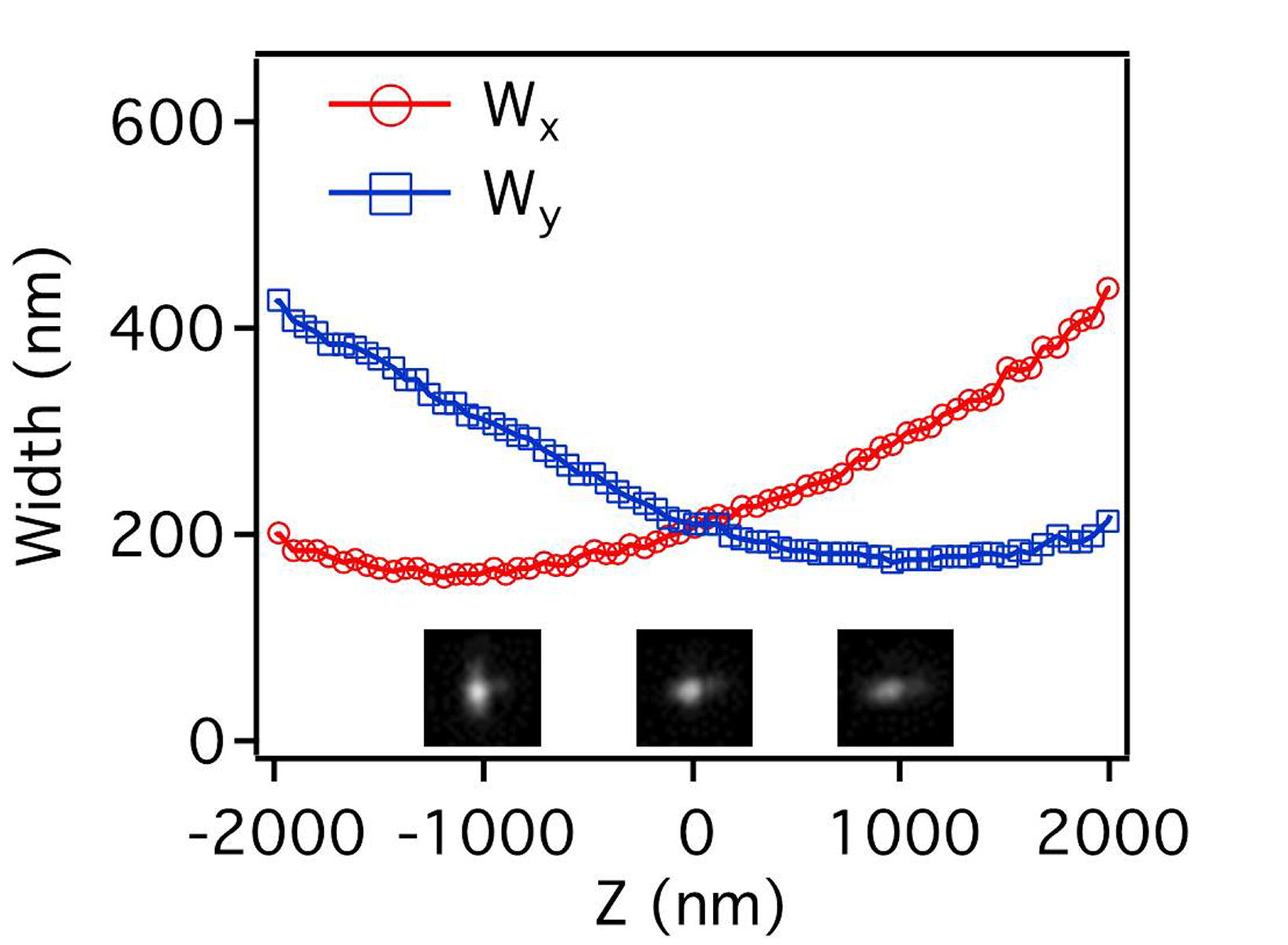 __ __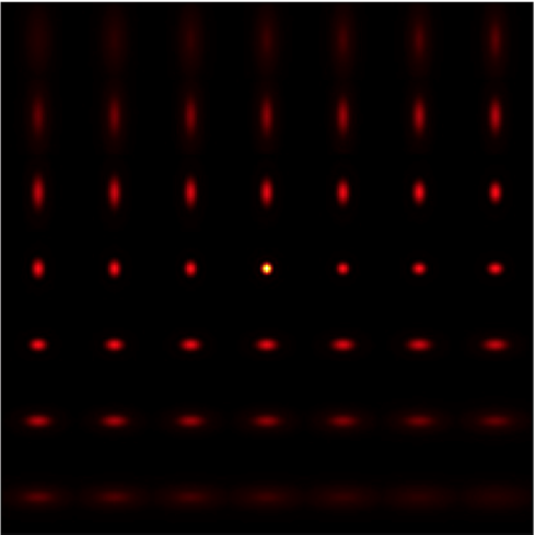
Fig. 2: (Left) Profiles of z-dependent PSF width and images of a fluorescent bead at three different z positions. The z-dependent curves of Wx and Wy were produced by fitting the images to an elliptical Gaussian function. (Right) Astigmatically distorted PSFs at different z and x positions are displayed. A data acquisition protocol depicted in Fig. 3 was developed for synchronously moving imaging focal plane with light sheet through a desired imaging volume. Assuming the scanning range is Zo, light-sheet thickness d=3um, depth-resolving range do=2 um, and exposure time of camera ts, the data taking time per scan is about ts(Zo/d)(d/do). The 3D localization can be accelerated to a significant factor with a mixed CPU/GPU implementation. 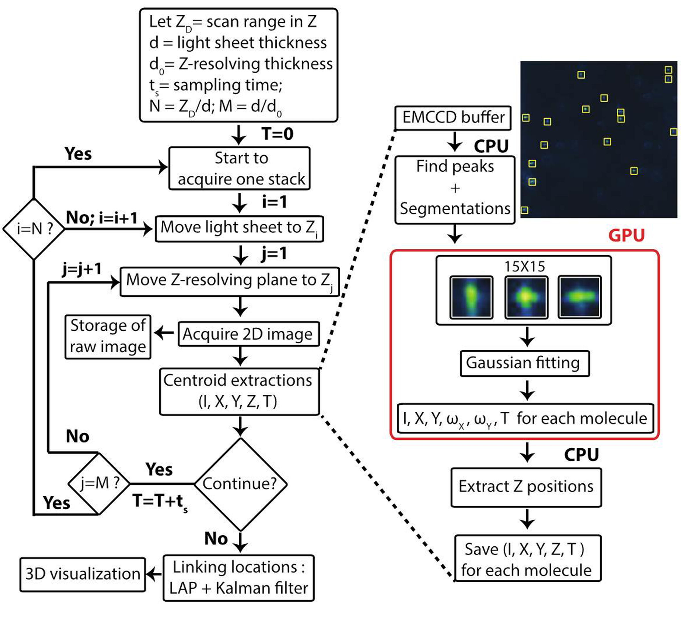
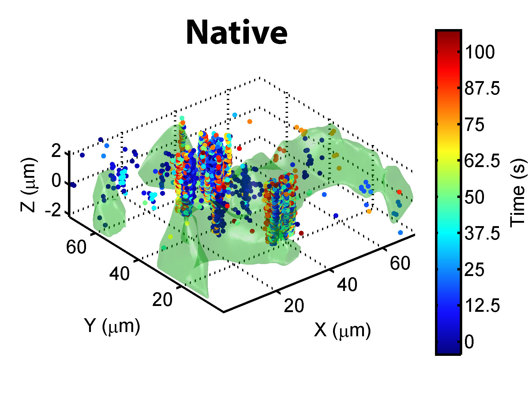
Fig. 3: Flow chart of the data acquisition protocol for single-molecule 3D localization with time-lapsed light-sheet microscopy. To verify the functionality, we applied the apparatus to investigate the translocating process of cell penetrating peptides (CPP) across the plasma membrane of live cells. Single-particle trajectories (SPT) can be constructed from time-lapsed images of the light-sheet microscope. However, connecting the acquired location coordinates to generate 3D trajectories is challenging with many difficulties. To circumvent the difficulties, we formulated SPT as a linear assignment problem (LAP), which can yield a globally optimal solution for multiple particle tracking. The goal of LAP is to find the assignment matrix between the measured location coordinates and predicted positions with minimal cost. The LAP can reliably predict the directed movements of particles. For diffusive motions, we implemented a Kalman filter as a diffusive movement predictor, which can provide an optimal estimate for the states of linear dynamic systems in the presence of Gaussian noise. By combining LAP with the Kalman predictor, we can reliably reconstruct 3D trajectories from the time-lapsed location data of the light-sheet microscope. Figure 4 presents an example of 3D trajectory reconstruction (lines) from coordinates (symbols with apearing time color coded) measured by the time-lapse scanning light-sheet microscope. 
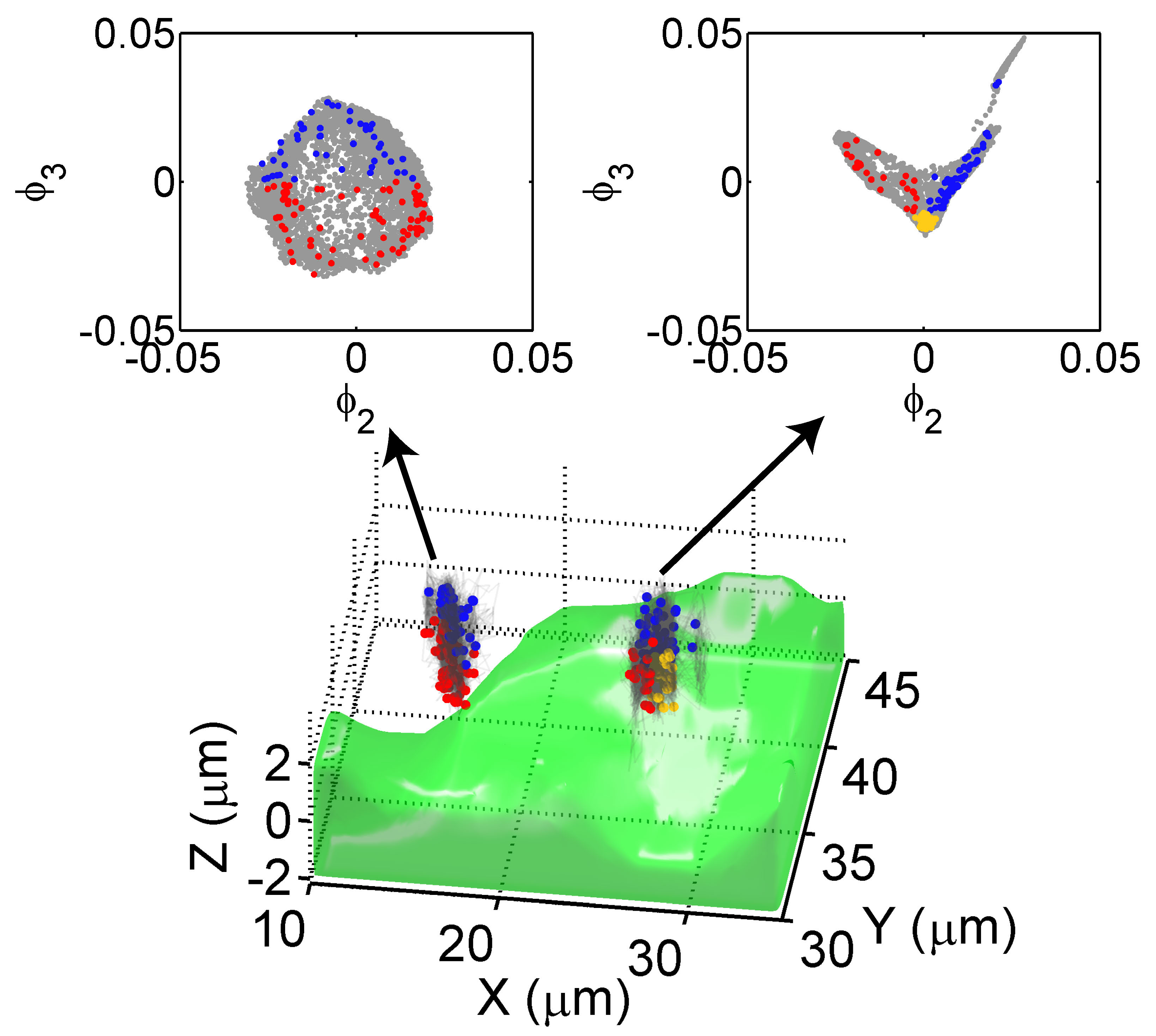
Fig. 4: 3D trajectory reconstruction (lines) from coordinates (symbols with appearing time color coded) measured by time-lapse scanning light-sheet microscope. Large numbers of single-molecule trajectories in live cells can be collected at tens-of-nanometers precision. Those acquired data are actually spatially and temporally coarse-grained from sampling of real molecule motions. Those sampled trajectories cannot be modeled by the overdamped Langevin equation. Therefore, to uncover local biophysical mechanism underlying molecule motion from those sampled trajectories, a stochastic model of the acquired data has to be developed. To gain insight into the dynamic process related to the observed molecules, a Markov state model from those trajectories was built to disclose successfully the diffusion states involved in the translocating process of CPP across the plasma membrane. (2) Femtosecond 2D Coherent Spectroscopy of Energy-Harnessing MaterialsThe collective excitation of Coulomb-coupled nano objects is an important phenomenon in energy harnessing materials. Examples can be found in biological light harvesting complexes, which consist of several chromophores, and an artificial system forming by metal nanoparticles/semiconductor quantum dots. Dipole-dipole coupling occurs on a nanometer scale, causing the states of the individual photo-emitters to hybridize and form new collective states, which delocalize over the whole structure. Long-lasting coherence has been discovered in photosynthetic complexes and attributed to from strong correlation between the protein-induced fluctuations in the transition energy of neighboring coupled chromophores. This long coherence enables highly efficient energy harvesting in photosynthesis. 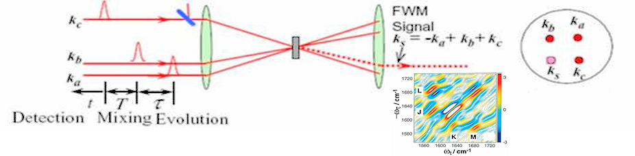 _ _

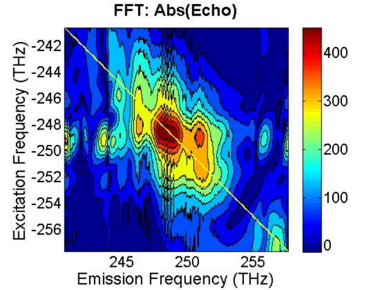
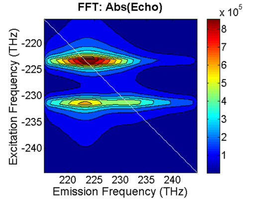
Two dimensional coherent spectroscopies (2D CS) can probe the couplings between optical excitation modes, and therefore allows for extraction of optical excitation dynamics. The parameters needed to characterize each individual quantum process pathway are the transition energy, dipole moment and relaxation rate, which can be extracted directly from 2D CS. The relaxation rates are the most difficult to determine because the population decay and coherence dephasing in a complex system can be affected by many factors, including inter-particle many-body interactions and coupling to the environment, which can substantially change the system dynamics. Moreover, isolating the relaxation rates associated with different quantum processes is often difficult. Lineshape analysis of 2D CS can overcome these difficulties to reveal the relaxation dynamics. Semiconductor quantum dots (QDs) are promising candidates for a number of optoelectronic applications. We applied 2D CS to investigate InAs quantum dots in near IR region. We observed cross peaks on the 2D plot, verifying the existence of coupling among the QD units. This technique can also be useful for other coupled systems for light harnessing applications. 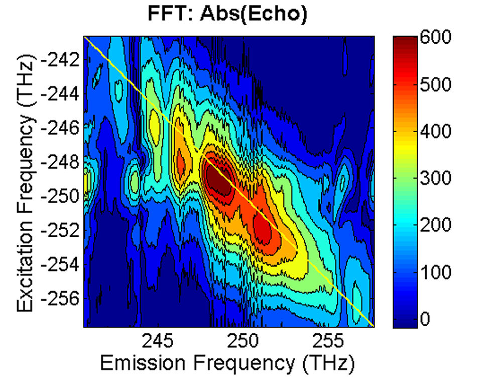
More....Write to the authors, press here (3) Single-Molecule BiophysicsOptics has become a valuable tool in life science. If an optical process is employed, the resulting technique can possess both single-molecule sensitivity and minimum invasiveness. Single-molecule techniques have matured into powerful tools for probing the complex behaviors of biological molecules. Catalysis-Associated Conformational ChangesIn single-molecule imaging, the distributions and fluctuations of important parameters can be obtained, yielding deep insights into the structure, the reactions, the transitions between molecular states, and the micro-environments surrounding the molecules under study. 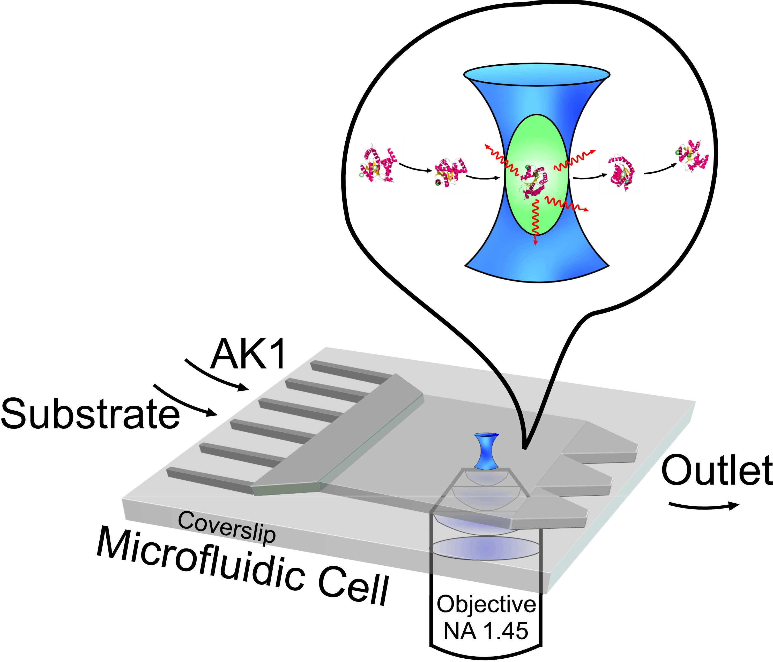 --- --- 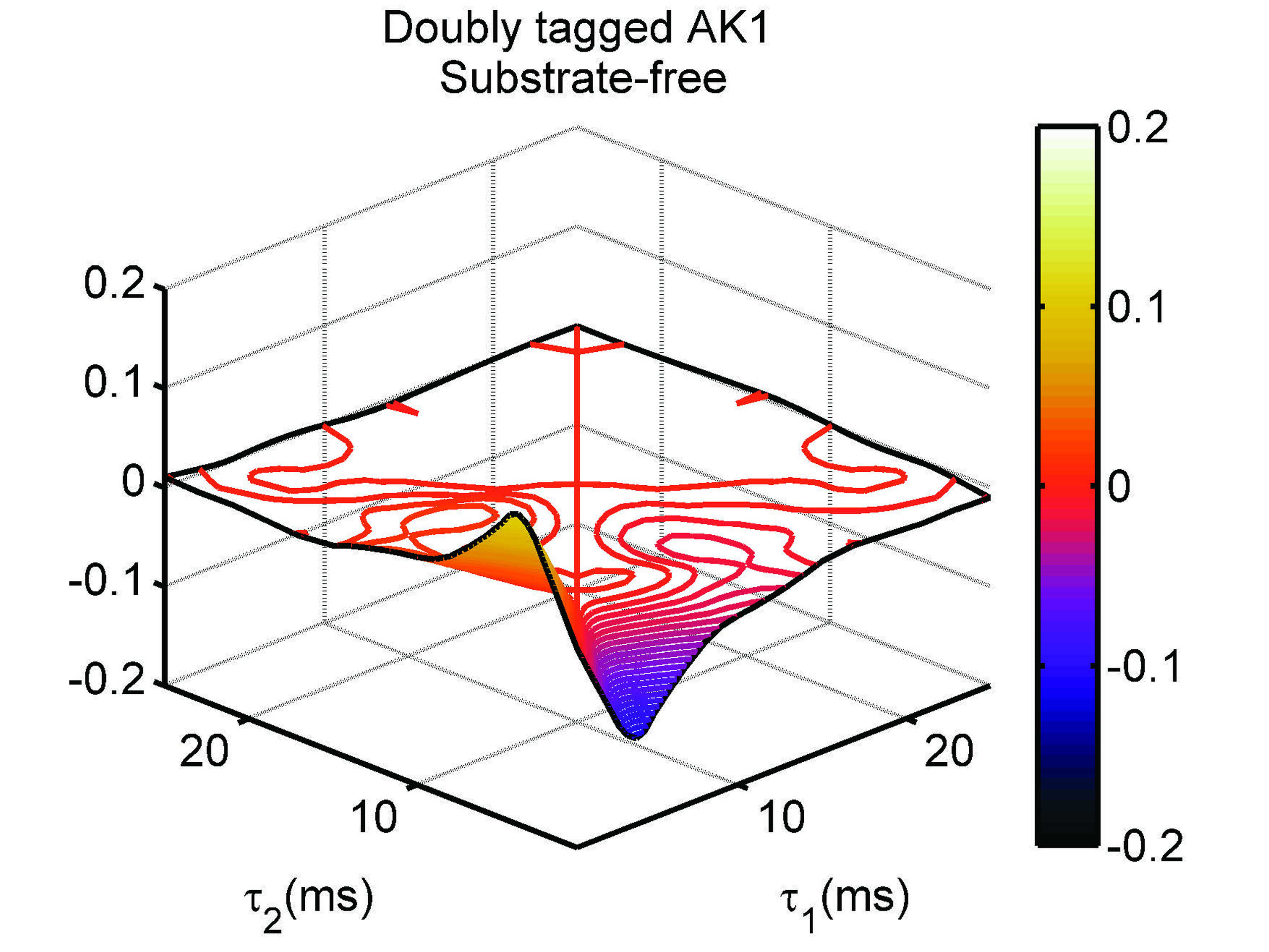
(Left) Optical microscope with microfluidic device employed for single-molecular detection and imaging, (Right) Difference of the second-order correlation functions reflects time-irreversability of a single-molecule adenylate kinase isoform 1 (AK1) in a nonequilibrium steady state. We tested this idea on the enzymatic action of human adenylate kinase 1 (AK1) at the single-molecule level. We labeled the core domain of AK1 with two identical fluorophores. Due to the self quenching effect, the conformation changes of the core domain are encoded into the fluorescent intensity fluctuations. We discovered the resulting second-order correlation functions of photon fluctuation traces do not possess time-reversal symmetry, indicating single-molecule AK1 is in a nonequilibrium steady state. More ... Signaling Machinery in Live Cells Studied by Single-Molecule OpticsThe EGFR can form homodimers with itself and heterodimers with other members of its receptor-family, such as ErbB-2. Previous study suggests that either dimers are preformed or EGF binds only to monomeric receptors that subsequently form dimers. ErbB-2 receptors are overproduced in some cancers and that overproduction can alter the dynamic behavior of the EGFR, presumably resulting from a shift of the normal distribution of EGFR homodimers to favor the EGFR–ErbB-2 heterodimers. This mechanism is poorly understood. We hope to decipher the mechanism to understand and manipulate receptor function at the molecular level. More ... 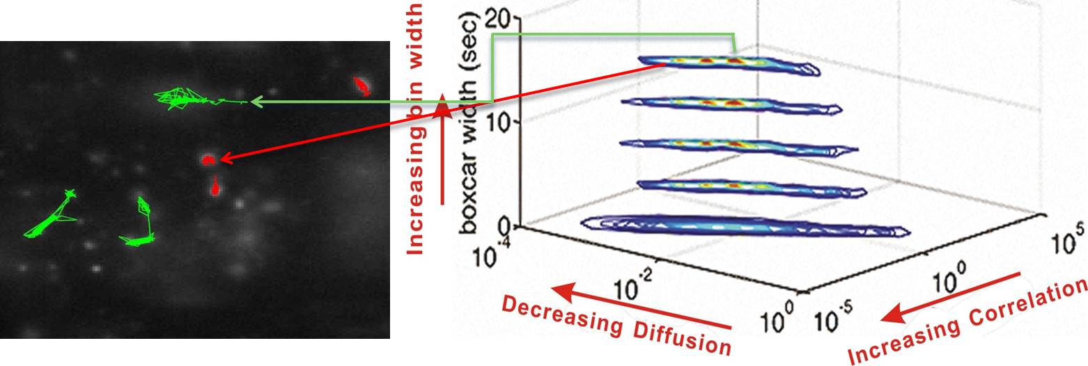
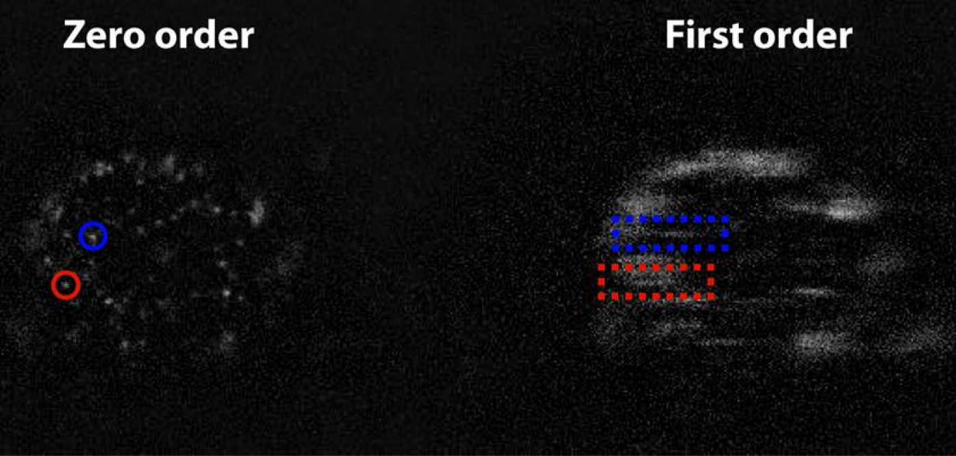
(Left) Fluorescent image of single-molecule trajectories of Ab-alexa 488-EGFR (green) and EGF-cy3-EGFR (red) on live He-La cells. (Right) A moment analysis of the single-molecule trajectories was developed for revealing the EGFR association mechanism. Simultaneous Tracking of Location and Spectrum of Fluorescently Tagged EGFR in Live CellsWe improved the single-molecule optical microscopy to simultaneously acquire the location and spectrum of a tagged protein in a live cell by placing a transmission grating before the imaging lens of the microscope. The zero-order image provides the location data with a spatial resolution of 30 nm; while the first-order diffracted image carries the spectral information. The following figure shows a typical result with the apparatus. Two cy3-taged EGFRs in a living He-La cell; one locates at the position marked by red circle (see the zero-order image), the other at the position emphasized by the blue circle. The spectrum emitted by the fluorophores forms a line in the first-order image emphasized by the red and blue rectangles, respectively. More ...
(a)
 ...(b) ...(b)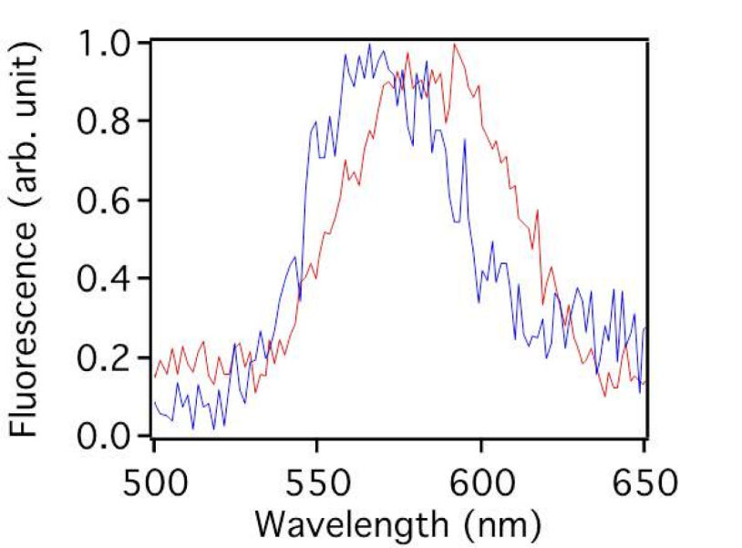
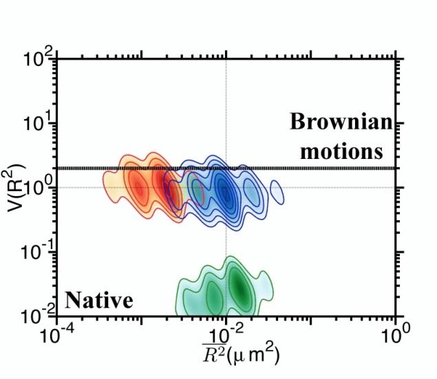
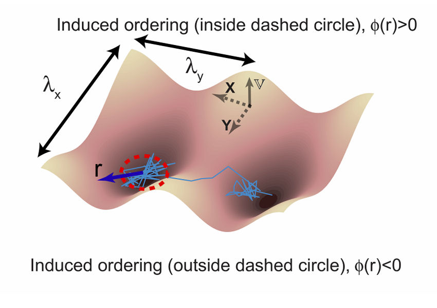
(a) Zero-order diffraction image and the corresponding first-order image of Cy3-ab-EGFRs in a live HeLa cell. (b) The fluorescent spectra converted from the line profileslying inside the blue and red dotted rectangles of (a). Energetic Model of Dynamic Regulation on Receptor Complexes by Actin Corrals and Lipid Raft DomainsWe developed an energetic model that captures both statistics of single-molecule trajectories and complex structures of plasma membranes of live cells. Simulation results on the diffusive trajectories of receptor proteins under varying confinements from actin corrals and lipid raft domains were presented in a plot of normalized variance V(R2) versus local mean-squared displacement R2. Our energetic model can serve as a useful platform to improve our understanding of how the mobile active clustering process of receptors and membrane structure regulates signal transduction. More ... 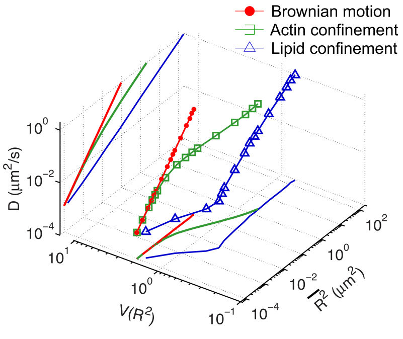
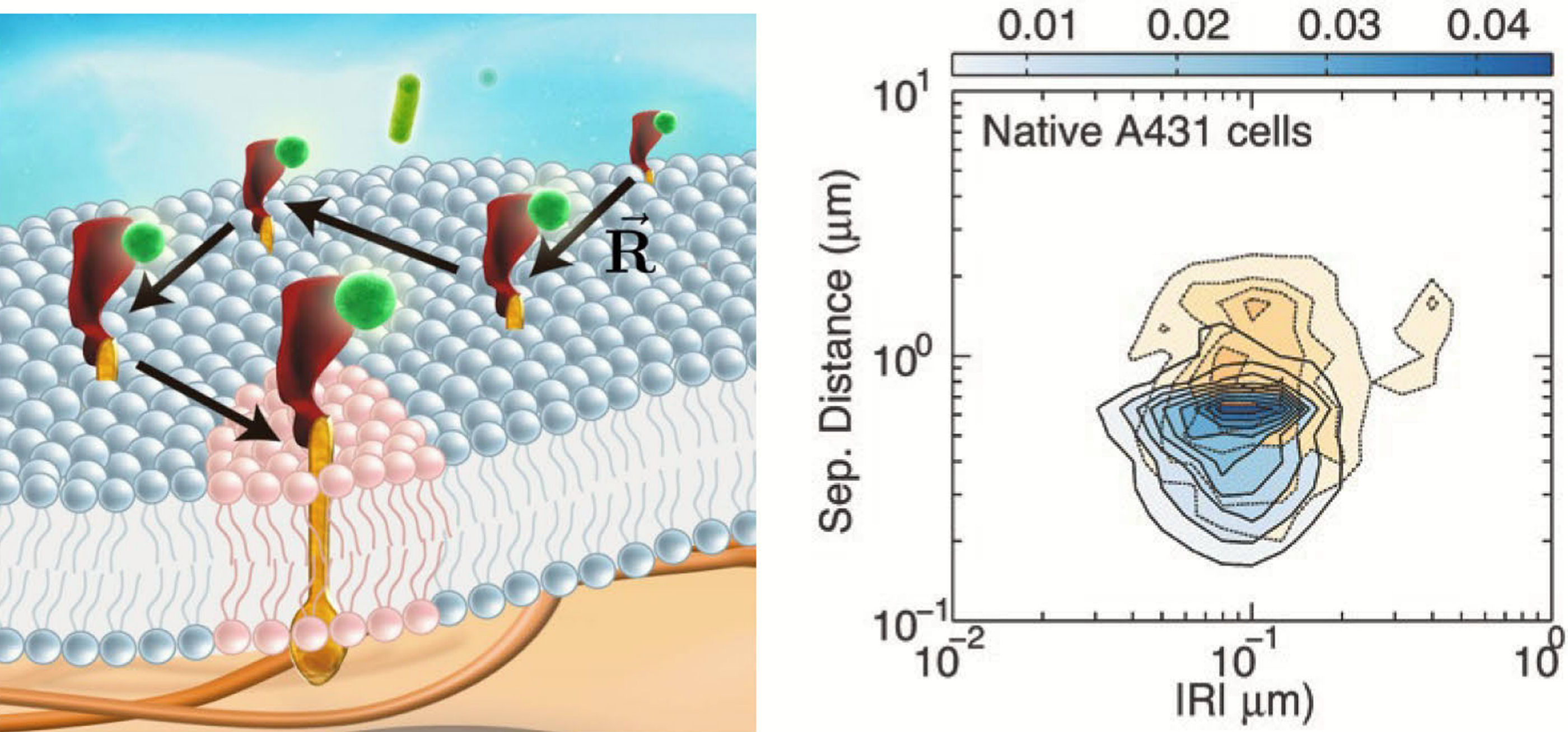
Simulation results of D-V(R2)-R2 for single-molecule receptors under Brownian motion (red solid circles) and diffusive motion under the confinement from actin corrals alone (green open squares) or both actin corrals and lipid raft domains (blue open triangles). Unraveling the Impact of Lipid Domains on the Dimerization Processes of EGFRs of Live CellsWe presented the measured single-molecule trajectories of EGFRs on live cells in the plot of V(R2) versus R2. An attractive feature of this data visualization scheme is that when a molecule repetitively visits a membrane domain, the characteristic R2 and V(R2) of the domain will be imposed on the trajectory; this yields a peak feature at the corresponding position on the plot. By exploiting the technique, we discovered that after ligand binding, EGFR molecules can move into lipid domains, whereas unliganded species remain outside the lipid domains. The lipid domains surrounding two liganded EGFRs can merge during their correlated motion. The conformation of the EGFRs in a dimer in a lipid domain exhibits a fairly heterogeneous distribution of distance between the two bound ligands. Our quantitative data analysis method captures dynamic receptor interactions at the single-molecule level, providing details that are often obscured in other methods. More ... 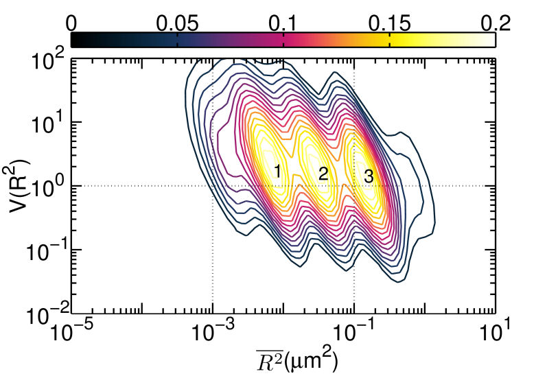
V(R2) versus R2 plot of unliganded Cy3-Ab-EGFR on live He-La cells at rest. |

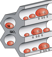
Research Projects(1) Architectural Photonics: assembling artificial functional materials from nano building blocks to enhance opto-electronic responsesArtificial functional materials self-assembled from nano building blocks offer great potential in many scientific disciplines and provide a modular solution for an increasing demand of multi-functional materials. Recently, we have been exploring several plausible ways to realize the concept of “architectural photonics” with improved opto-electronic responses by tailoring device geometry or structure of material. Specifically, we have realized some useful materials with interacting subunits combined in a directed hierarchically organized structure based on a preferential growth technique of silicon quantum dots in the pore-channels of mesoporous silica. We have demonstrated enhanced NIR photodetection, non-volatile memory, and photovoltaic conversion with the artificial material system. 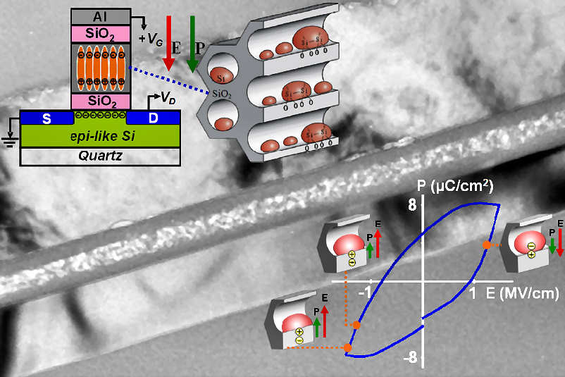
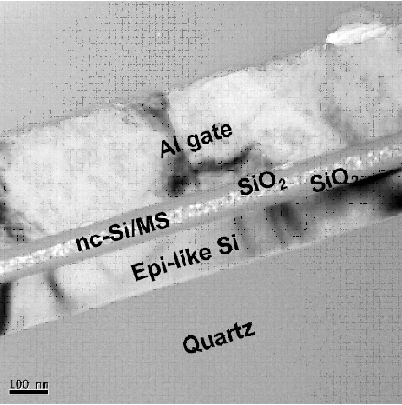
Schematic of an artificially engineered optoelectronic material by preferentially growing silicon quantum dots on the bottom of the pore-channels of mesoporous silica. A thin layer of the material had been integrated into the gate oxide stack of a MOSFET to demonstrate the functionality of nonvolatile memory. Several advantages of using quantum dots in making artificial crystals. Firstly, the wide choice of lattice structure and secondly interdot coupling and the properties of quantum dots can be modified separately. Other applications of this technique include enhanced charge transport in CdSe quantum-dots or nano rods embedded copolymers, and enhanced photoluminiscent emission from TiO2 nanocrystals or CdS QDs-embedded conjugate polymers. 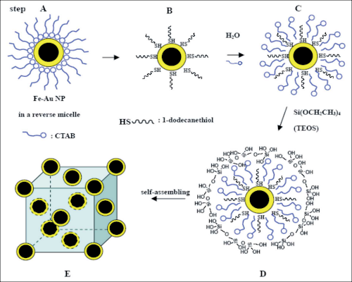 ---- ---- 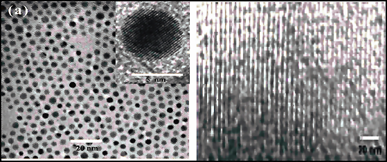
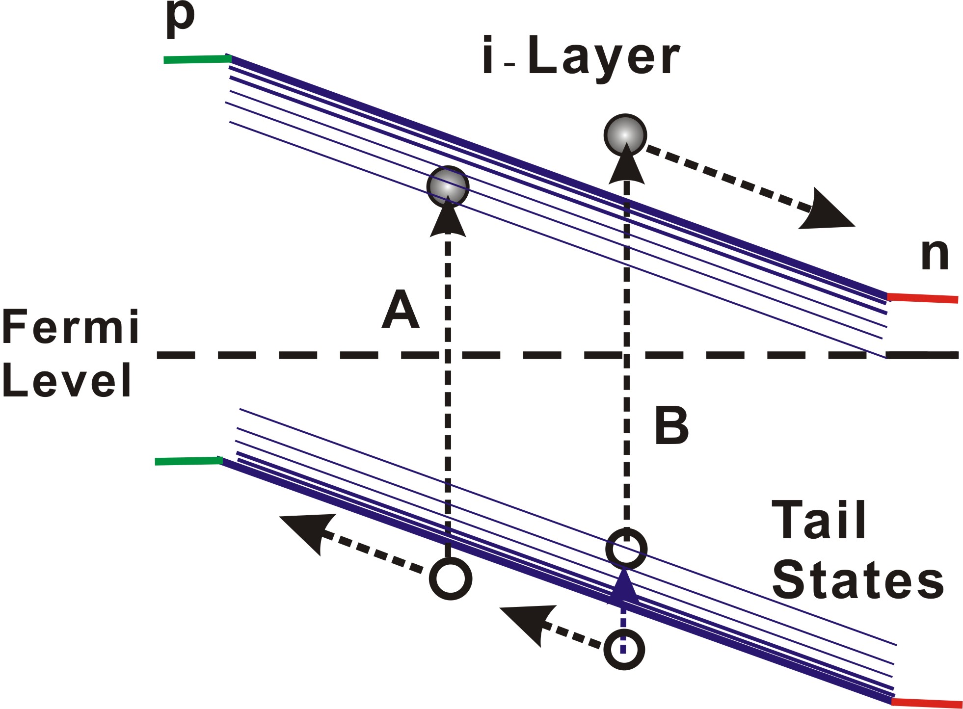
A versatile scheme (left) can be used to self assemble a variety of nano building blocks (middle) into 3D artificial crystals (right). (2) Developing Low-Cost Technology for High-efficiency Thin Film Solar CellsPhotovoltaic application requires further scientific breakthroughs in order to achieve the planned target in 2030. To help advancing toward this target, we focus on the development of high-efficiency inexpensive thin film solar cell with inductively coupled plasma (ICP) deposition technique. High quality amorphorous hydrogenated silicon (a-Si:H) films were deposited at a fairly low deposition temperature of 140C. Single- and stacked pin solar cells with 400-nm thick i-layer had achieved a conversion efficiency of 9.6% with an improved light-soaking stability. (see Fig. 4). 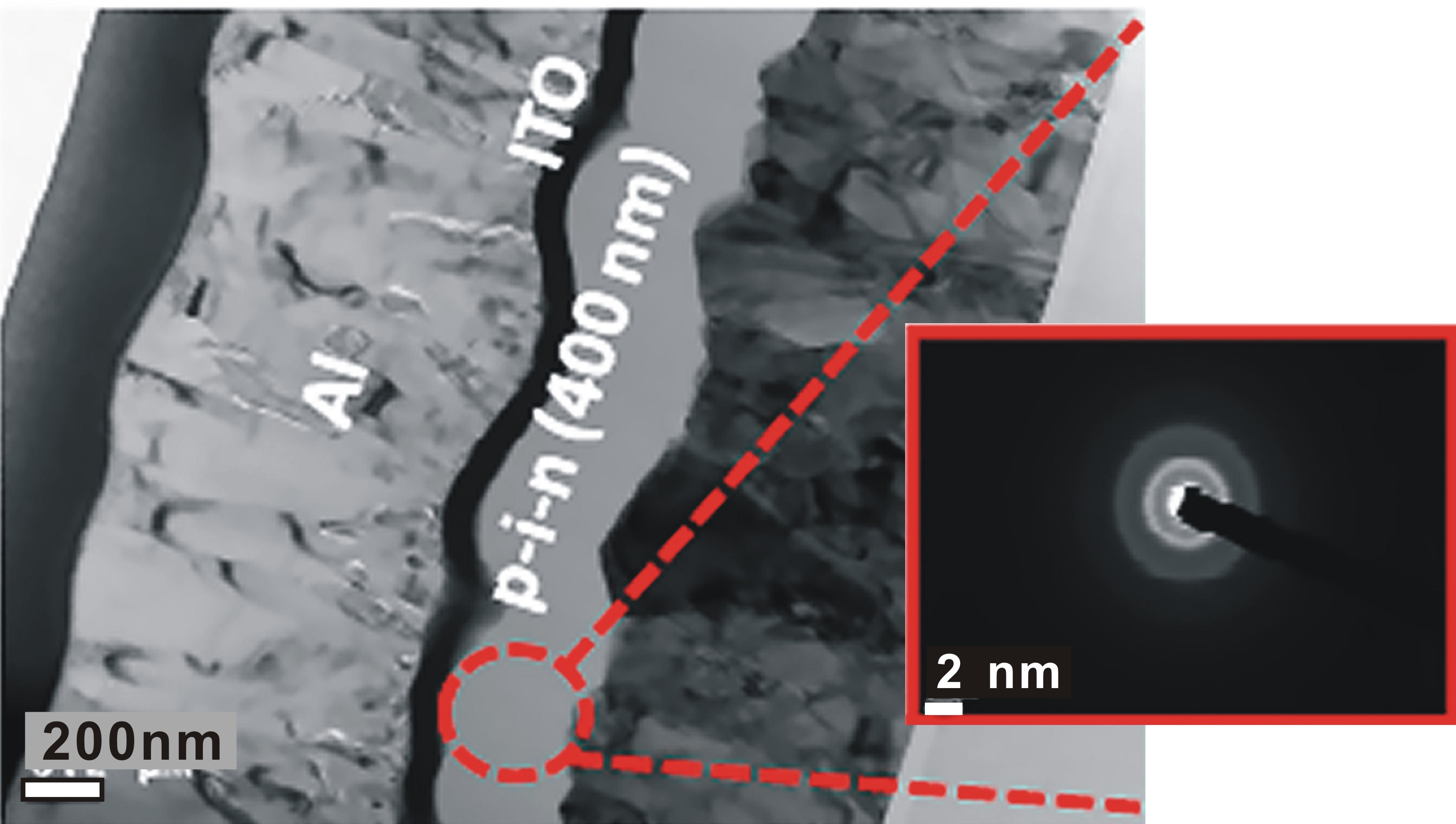 --- ---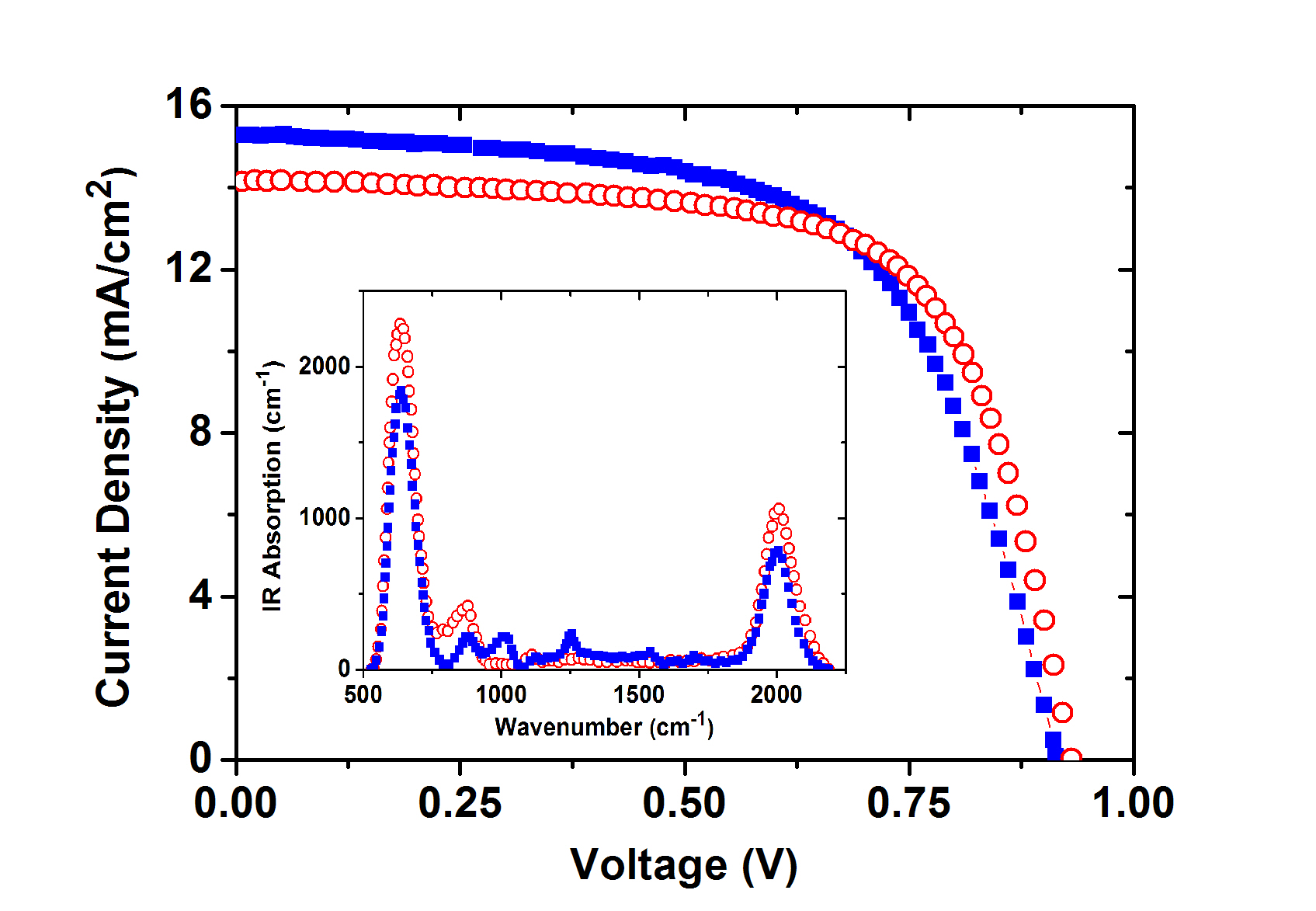
(Left) The cross-sectional TEM image showing the a-Si pin solar cell fabricated by ICPCVD on Asahi U substrate. Spatially resolved diffraction pattern of the a-Si pin stack reveals clearly the amorphous phase. (Right) J–V characteristic curves of a-Si pin solar cells deposited by ICP-CVD (blue solid squares) and VHF-PECVD (red open circles) at 140 C. Inset showing the FTIR spectra of 400-nm thick intrinsic a-Si layers deposited by the same methods. (3) Tracking correlative motion of moieties in molecular systemsTracking correlated motion of sub molecular fragments is crucial for understanding responses of complex molecular systems. We had developed time-resolved two-dimensional infrared (2D IR) technique and exploited it to investigate the field-induced reorientation dynamics of ferroelectric liquid crystal (FLC). We were able to reveal the detailed correlated motions of submolecular fragments of the FLC films. The important feature of the time-resolved asynchronous scan FTIR technique is that the achievable temporal resolution is limited only by the IR pulsewidth used. Therefore, if we can use the state-of-the-art techniques of laser and nonlinear optics to generate coherent light pulses with wide bandwidth and short duration, we shall be able to follow the dynamics of DNA-protein interaction and protein folding with the time-resolved asynchronous scan 2D IR technique. 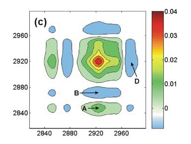 --- --- 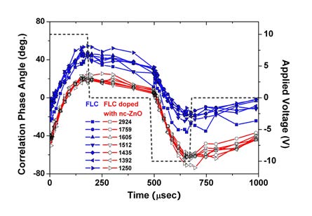
(Left) 2D IR synchronous correlation plot from the alkyl-chain modes of a surface stabilized ferroelectric liquid crystal (SSFLC). (Right) Time courses of 2D IR correlation phase angles of IR-active molecular normal modes of SSFLC with (open symbols) or without (filled symbols) nc-ZnO doping. The dashed line denotes the bipolar square wave form of applied electric field used. (4) Characterization of nanostructured material with advanced optical techniquesSpectral imaging of materials/devices with quantum controlThe purpose of femtosecond quantum-control study is not only to control the evolution of a complex quantum system but also to deduce the detailed dynamic mechanism from the optimal laser field used. Recently, we have developed a freezing phase algorithm appropriate for adaptive quantum control with an femtosecond pulse shaper. With the technique the desirable optical field characteristics can be verified and be used to selectively excite one type of probe molecule while leave the others in their ground state. Quantum control also offers an additional degree of freedom to distinguish coherent and incoherent nonlinear optical processes involved in a complex optical excitation. We had combined a scanning optical microscope with the femtosecond pulse shaping apparatus to yield the spatial distribution of a specific species with quantum control methodology. (see Fig. 6). 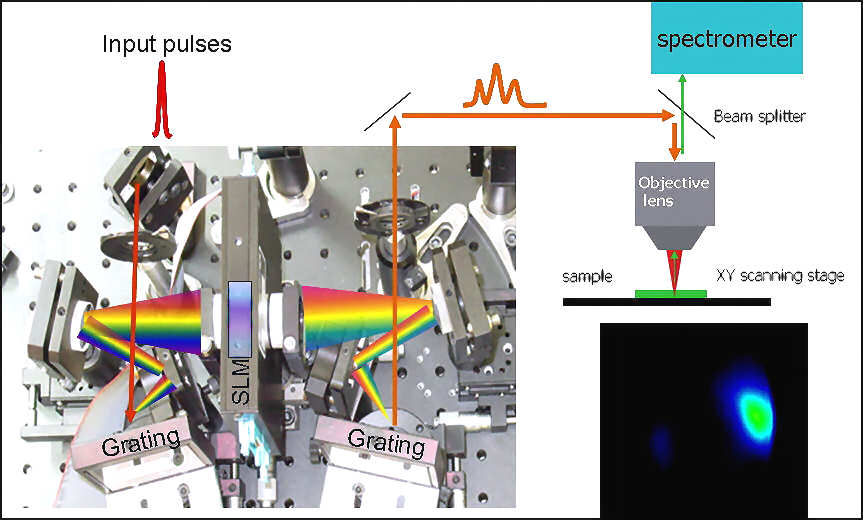
Apparatus can be used for molecular specific mapping with ultrafast coherent control methodology. To analyze extremely weak light-material interaction in real time, we also developed for the first time a single-shot OPA-FROG, which can be used to completely characterize a coherent optical field as weak as several atto joules. Complete-field characterization of advanced photonic materials and devices at the nanometer scales is also undertaking by combining scanning near-field optical microscopy (SNOM) with heterodyne interferometry. Inverse-problem technique for advanced liquid crystal DevicesRetrieving model parameters of material and device from measured data is useful while difficult task in view of the ill-posed nature of the inverse problem. We had developed a mathematical inverse problem approach to retrieve liquid crystal (LC) director profiles from optical transmittance measurements. We proposed a new regularization matrix based on a priori knowledge of LC. The method can reliably yield a LC director profile with noisy optical transmittance data in a finite range of incident angle. 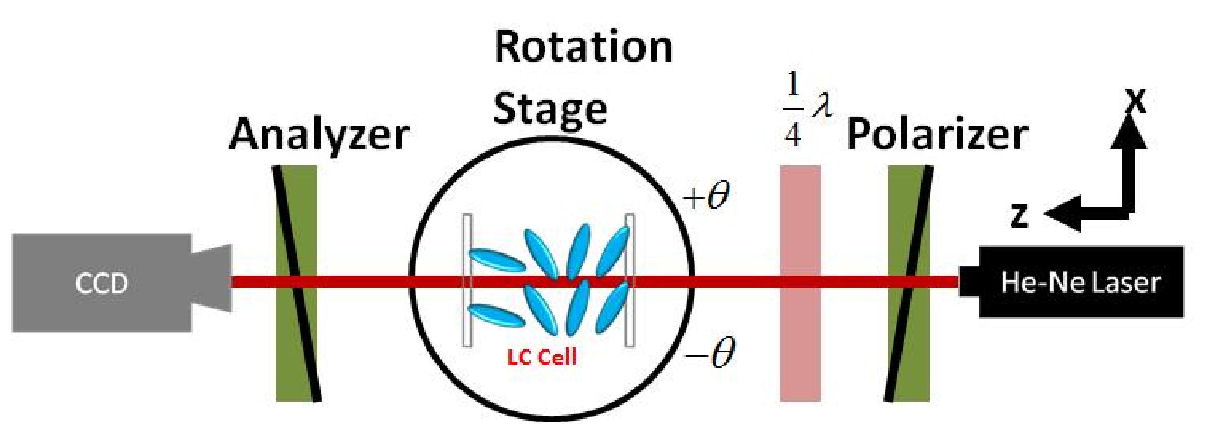
Experimental setup used to measure the optical transmittance data for inverse problem retrieval of LC director profile. We demonstrated that the functionality by retrieving the liquid crystal director profiles of differently aligned LC cells from experimentally measured data, indicating the method to be useful for the design of LC devices where the LC director profile needed to be carefully tailored. 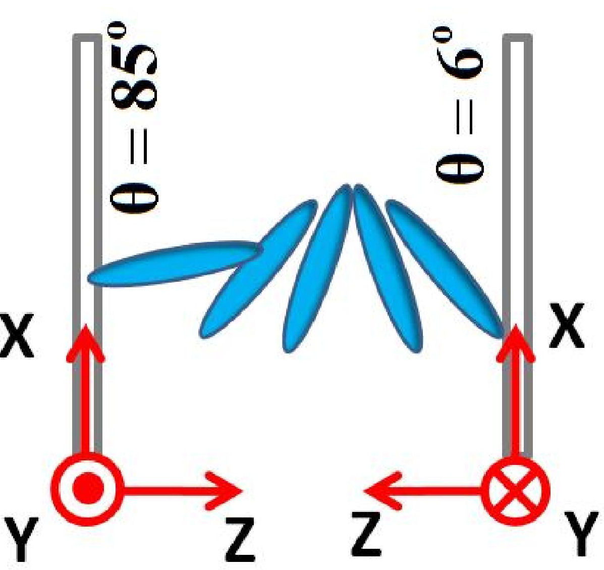 ----- -----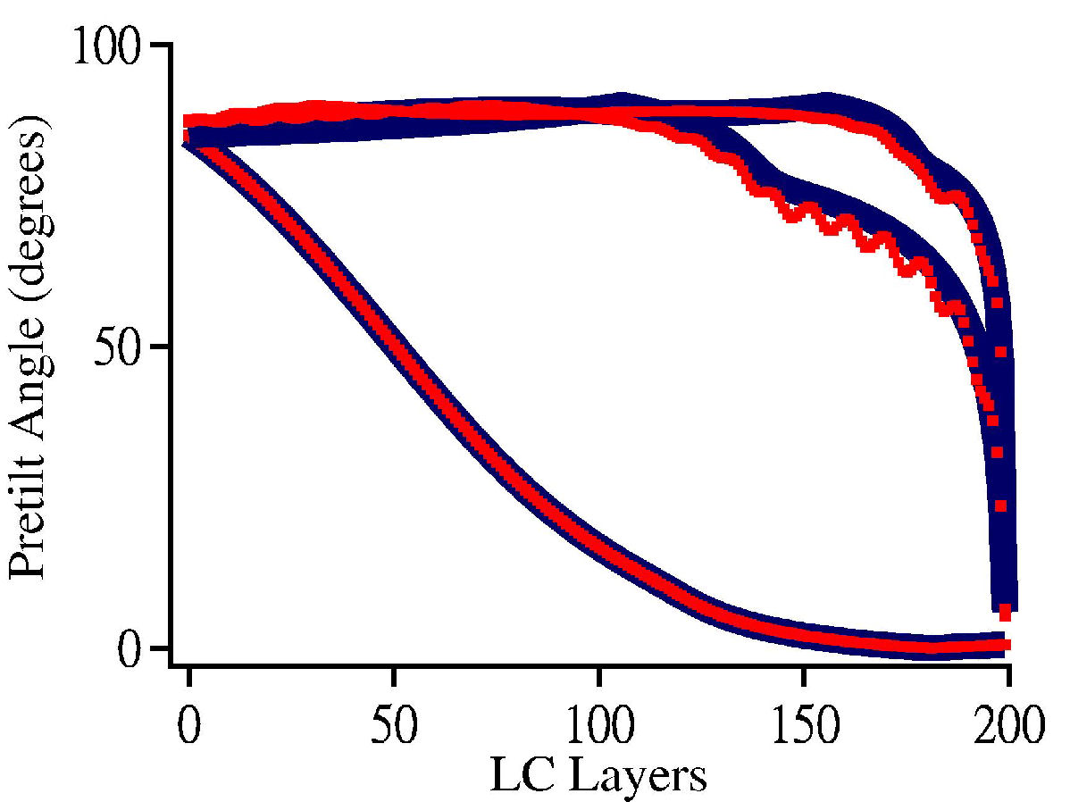
The retrieval director profiles of a hybrid cell by our inverse problem retrieval method. (Left) The coordinate system used to present the LC director profiles. (Right) The retrieved director profiles of the hybrid cell biased at 0V, 2.5V, and 5V. Two profiles are included for comparison: the curves of red filled squares: retrieved profile inversely from optical transmittance measurements, and blue solid curves: the simulated profile calculated by PDE solver with FEM and Q-tensor approach. Material Characterization with THz RadiationA cutting-edge analytical tool to probing the structural dynamics of biomolecular systems is a time-resolved two-dimensional infrared spectroscopy in THz region. With this technique, we can extract useful dynamic information about how biomolecules perform their biochemical function. However, to achieve this goal, we have to generate short THz radiation pulses with sufficiently high pulse energy for pump-probe type measurement. We had experimentally demonstrated the generation of single-cycle high-power terahertz radiation with two-stage optical rectification in GaSe crystals. By adjusting the time delay between the pump pulses employed to excite the two stages, the terahertz radiation from the second GaSe crystal can constructively superpose with the terahertz field injected from the first stage. The high mutual coherence between the two terahertz radiation fields is ensured with the coherent optical rectification process and can be further used to synthesize a desired spectral profile of coherent THz radiation for the applications of two-dimensional correlation spectroscopy. Probing optical response of a dense array of silver nanorods with light scatteringIntra- and inter-particle coupling effects are important but have not been taken into account properly to model the optical response of a dense array of nano objects. We present a method to analyze the optical response of a layer of tilted silver nanorods fabricated with oblique-angle deposition (OAD) technique. Our technique can render nonlocally coupled nano objects into an array of independent quasi particles. The effective polarizability tensor of the tilted silver nanorods retrieved with this method exhibits non-zero off-diagonal components, revealing information about the array structure and inter-particle quadrupolar coupling effect. Our technique is useful not only for the design of a photonic nanostructure with a specific property but also to yield insight into the correlation between the function and structure of optical metamaterial. 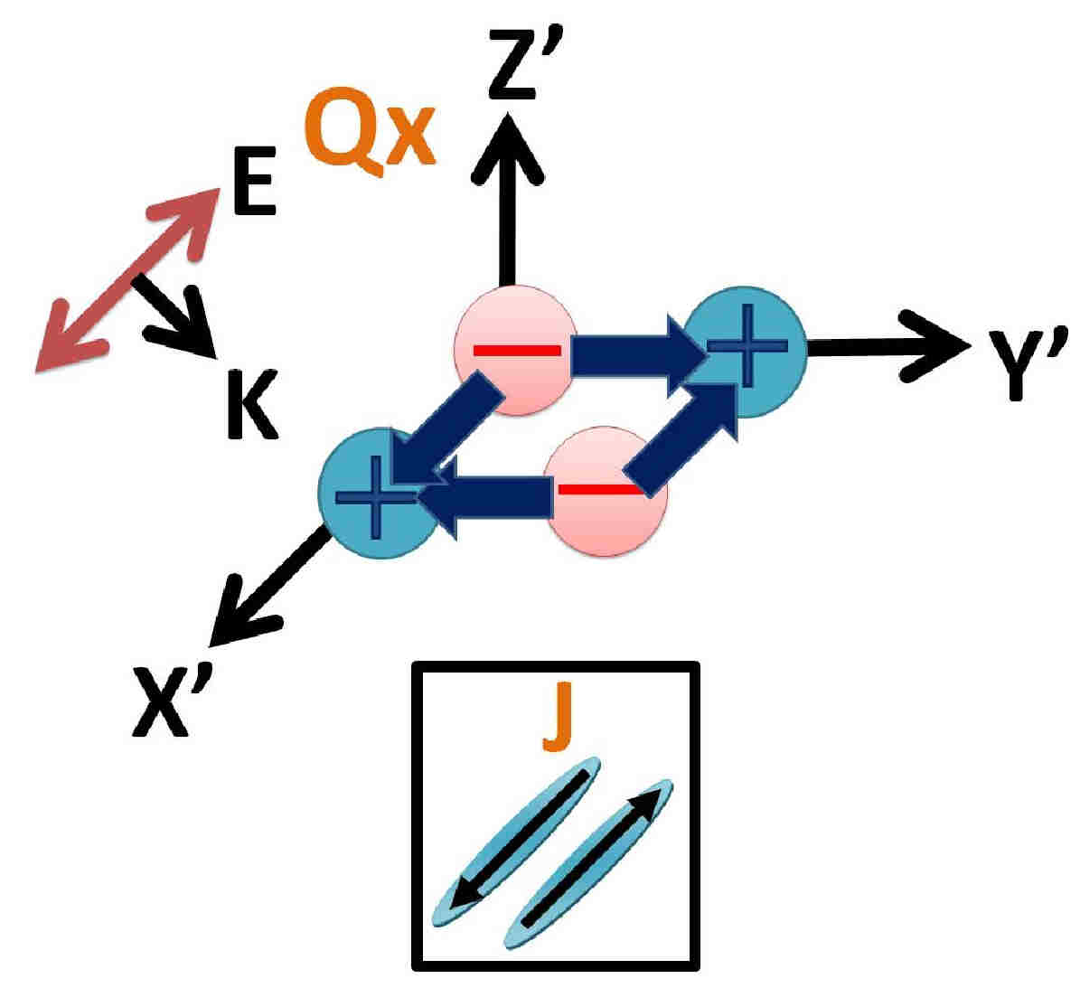 --- ---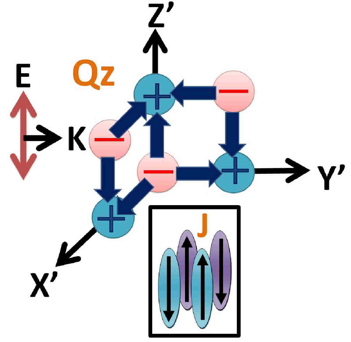 --- --- 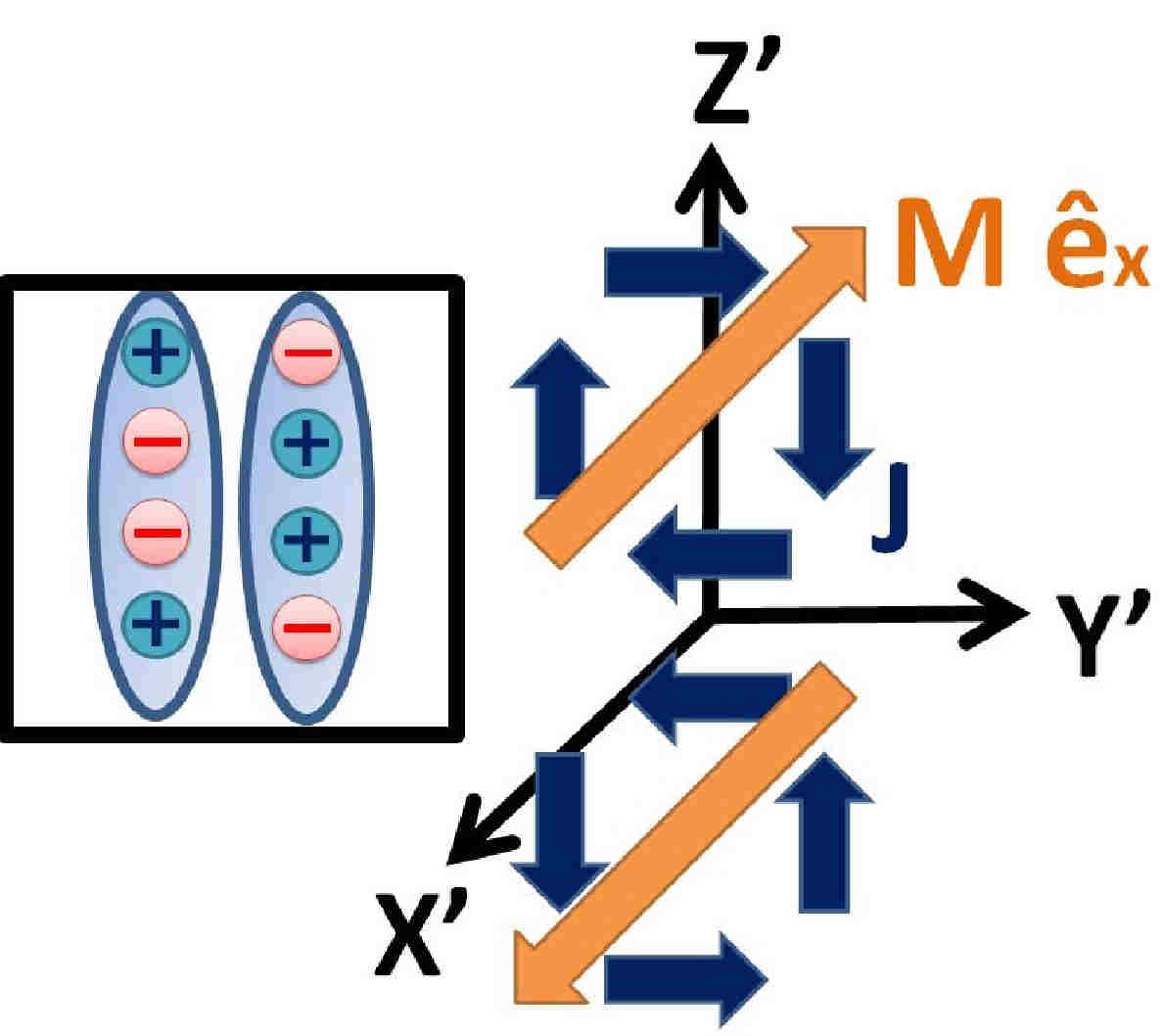
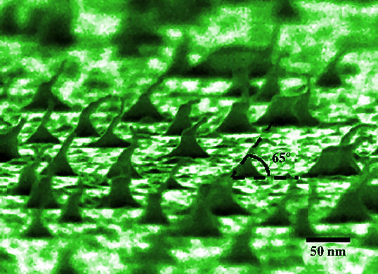
(Left) and (Middle) Schematic showing the asymmetric electron oscillation modes induced by an incident optical field with polarization along the principal axis of silver nanorods array. The arrows and ”+” and ”-” signs are the current and charge distributions, respectively. (Right) Illustrating the counter effect of magnetic dipoles from two neighboring nanorods. A monolayer of cyano biphenyl (8CB) liquid crystal (LC) molecules was deposited on an OAD substrate to probe the adsorbed geometries of molecules on the tilted silver nanorods. Our results show that the 8CB molecules are aligned along the Ag nanorod direction, and the main contribution of surface enhanced Raman scattering (SERS) is from the absorbed molecules on the side wall of the Ag nanorods. The information is useful for optimizing the performance of a sensing device where alignment structure of interfacial molecules plays a crucial role. 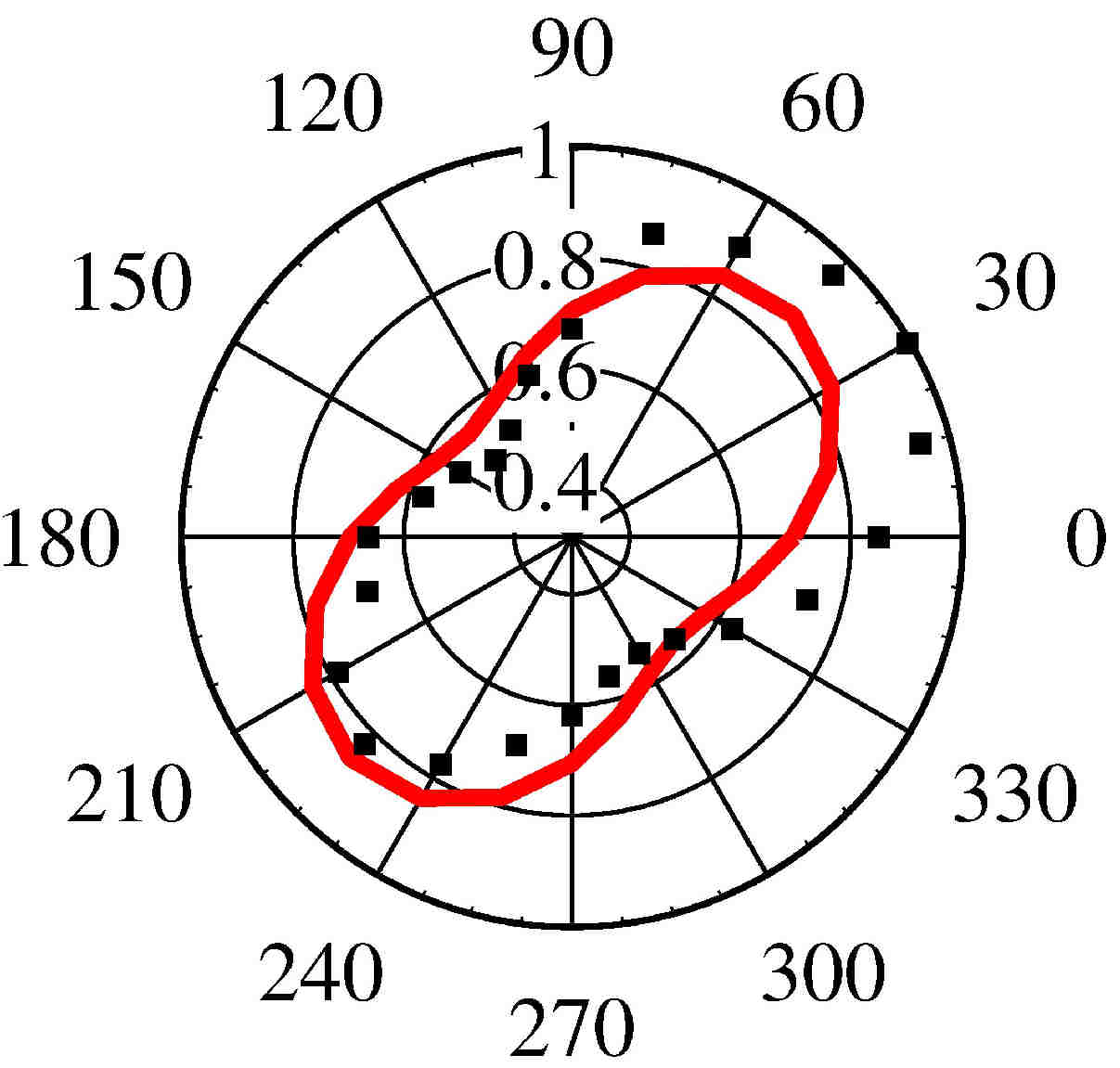 ----- ----- 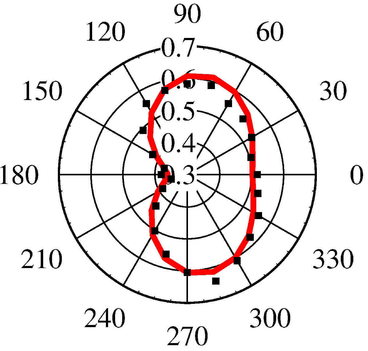
The azimuthal patterns of the inelastic Mie scattering from tilted Ag nanorods with a frequency shift of 800 cm−1 were measured (filled squares). The fittings to the model are presented in red solid curves. The zero azimuthal angle implies the tilt plane of the silver nanorods to be along the incident plane of the excitation light. (Left) The p-polarized scattering light was measured with s-polarized incident light. (Right) Both of the excitation and scattering fields are p-polarized. (5) Sum-Frequency Vibrational Spectroscopy of Liquid Crystal Molecules on Various Alignment SurfacesIn nearly all electro-optic applications of liquid crystals (LC), it is necessary to uniformly align the liquid crystal molecules to along a certain direction. Aligned polar structure of liquid crystal can be formed in a surface-stabilized ferroelectric liquid crystal (SSFLC) film or at an interface of a liquid crystal and alignment layer. Polar structure of SSFLC can yield a nonvanishing dipole moment per unit volume Ps, which is switchable with an electric field E via an interactive energy of -Ps.E. The resulting switching speed of SSFLC is much higher than that with nematic LC. Owing to the intrinsic surface and polar sensitivity, second-order nonlinear optical techniques such as second-harmonic generation (SHG) and sum-frequency vibrational spectroscopy (SFVS) had been widely used to investigate the structure of liquid crystal molecules on various alignment surfaces. To yield a clearer picture of the physical mechanism of LC alignment by polymer surface, an investigation of LC aligned structure on a number of different polymeric films with known surface structures is highly desirable. The latter information is often difficult to be obtained with SHG measurement alone. In this project, we studied the surface molecular structures of alignment layers treated by ion beams with SFVS. Imaging polarimetry and two-dimensional anchoring energy characterization were then used to reveal the anchoring strength, alignment quality and stability of LC film. More... 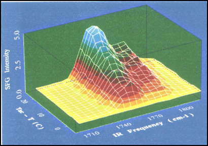
Sum-frequency vibrational spectra of SSFLC sample at the C=O stretch region are plotted as a function of the temperature of the sample. (6) Photonic System on Chip (PSoC)This multi-years project was supported by academic R&D program from Ministry Office of Economic Administration (MOEA). The first-phase research has two major targets: (1) develop the needed MOEM platform to realize a prototype silicon microbench blue-light optical pickup head, and (2) develop sub optical wavelength components and modules to facilitate further miniturization of future chip-scale photonic systems. The R&D team is formed by 19 PIs from various research centers and universities in Taiwan. During the first-phase investigation, the team members had published 171 referred journal papers, 149 conference papers, and filed 23 patents with 6 being granted. 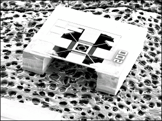
Actuator for Integrated Optical Pick Up Head |
Highlights – recent results
|