|
|
|
The Laboratory of Optical Studies for Advanced Materials (LOSAM) endeavors to develop and characterize frontier materials with novel laser techniques. To pursue this goal, the laboratory is equipped with (1) high-power femtosecond laser facility, (2) widely tunable picosecond coherent light sources; and (3) Yb-doped fiber laser pumped femtosecond laser with pulse shaping apparatus, etc. In this web page, I present a brief introduction of our research with sum-frequency vibrational spectroscopy of polymeric surfaces. The information was initially prepared for a discussion forum of my group. However, I will appreciate any feedback you are willing to give. |
Probing the Nanostructure of Co-Polymeric film with SFVSBy adjusting the hydrophobic/hydrophilic natures of polymer segments, block copolymers can self assemble into a film with a variety of ordered structures at the nanometer scales. To advance our knowledge of the interfacial bonding nature of block copolymers, we can apply infrared-visible sum-frequency vibrational spectroscopy (SFVS) to investigate this type of material. SFVS belongs to the class of second-order nonlinear optical processes and is known for its unique capabilities in measuring surface sensitivity and molecular specificity. 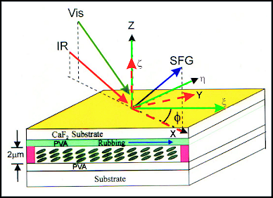
FIG. 1. Optical setup of SFVS for studying structures and orientations of molecules on varying alignment surfaces. Here we present an example of SFVS study on a film of PS-PMMA copolymer. The as-deposited film exhibits a lamina (layer) structure at the nanometer scale. The ssp SFVS reveals a polar structure of C-H bonding at interfaces or on the film surface. After annealing, the polar layer at surface and nanostructures in bulk vanish to form a homogeneous film, as indicated by the disappearance of the C-H stretching signature. However, the annealed film may still possess closely coupled dipolar pairs (i.e., the quadrupolar terms) spacing at a length scale comparable to the optical wavelength, causing significant nonresonant sum-frequency susceptibility. 
FIG. 2. ssp-Sum frequency vibrational spectra of as deposited PS-PMMA copolymer film and annealed film at the C-H stretch region. |
Modification of Polymeric Surfaces with Ion BeamsAs most polymers do not posses required surface properties for industrial applications, different kind of surface modification techniques were applied. One of the most versatile techniques is plasma treatment, which could produce unique surface properties while not affecting polymer bulk. Treatment of polymers with an inert gas (such as Ar) plasma leads to the formation of free radicals but does not add new chemical functionalities from the gas phase. Functionalization, for example, incorporation of oxygen, occurs after the treated sample was exposed to atmosphere or other environment containing oxygen. PS treatment up to an ion dose of 1e13 cm-2 shows reduction in surface aromaticity and broadening in the main carbon peak, which could be due to the formation of the new bonds. Here are some SFVS studies to reveal the ion-beam effects on polymeric surfaces. A. Influence of the angle of incidence of H-ion beam ---> Higher AOI resulting in higher de-aromaticity effect 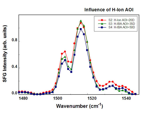
FIG. 3. ssp-Sum frequency vibrational spectra of SE7492 samples at the C=C stretch region. The samples were treated with H-ion beam with a fixed ion-exposure 5´1015 and incident angle varying from 20 degrees to 50 degrees. B. Influence of H-ion dosage Higher flux yields higher de-aromaticity effect on phenyl ring 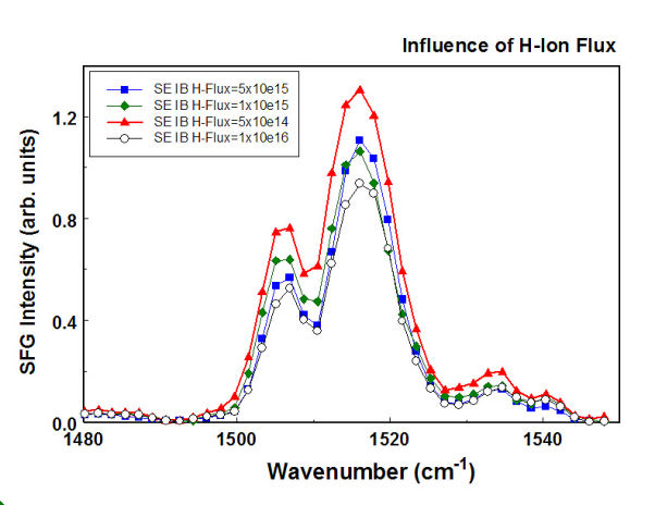
FIG. 4. ssp-Sum frequency vibrational spectra of SE7492 samples at the C=C stretch region. The samples were treated by using an H-ion beam with a fixed incident angle of 35 degrees, while the ion dosage was varied from 5 e14 to 1 e16. C. Influence of the kinetic energy of H-ions Higher kinetic energy of ions gives higher de-aromaticity effect on phenyl rings 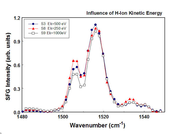
FIG. 5. ssp-Sum frequency vibrational spectra of SE7492 samples at the C=C stretch region. The samples were treated by using an H-ion beam with a fixed incident angle of 35degrees and the H-ion exposure 5e15, while the kinetic energy of the H-ions was varied from 250 eV to 1000 eV. D. Influence of ion species Argon ions yield higher de-aromaticity effect on phenyl rings, resulting in disappearance of C=C stretching (see open squares). However, the resulting radicals will re-form chemical bonds with other species after exposing to air, as shown by the recovery of SFVS spectrum after argon IB treatment (not shown in this figure). 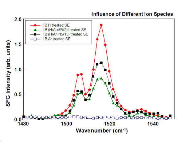
FIG. 6. ssp-Sum frequency vibrational spectra of SE7492 samples at the C=C stretch region. The samples were treated by using an ion beam with a fixed incident angle of 35 degrees, ion exposure 5e15, and kinetic energy 500 eV, while the ion species were varied by controlling the input partial pressure ratio from pure H2, H2/Ar=99/2, H2/Ar=15/15 to pure Argon.  __ __ 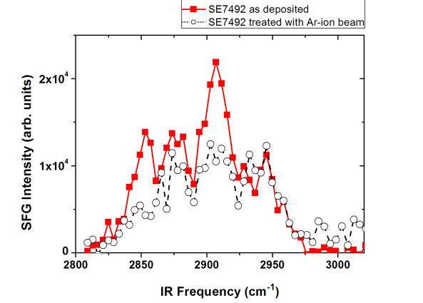
FIG. 7. ssp-Sum frequency vibrational spectra of SE7492 samples at the C-H stretch region. The samples shown here are untreated sample (filled squares) and the ones treated with pure H-ion beam (Left), and treated with the pure argon-ion beam (Right). The ion beams used have the following parameters: a fixed AOI angle of 35o, ion dosage of 5´1015, and kinetic energy 500 eV. Re-bonding of radicals can occur at the same time of H-ion beam treatment, but not by using argon ions. Only pure damaging effect was observed with argon ion beam. |
Surface Alignment ControlThis study indicated that a liquid crystal polymer (LCP) self assembled on a photo-irradiated surface could be used to yield surface alignment control. Sum-frequency vibrational spectroscopy revealed that the phenyl groups are aligned epitaxially, yielding an in-plane alignment order much higher than that at the photo-irradiated surface. Experimental Geometry of SFVS 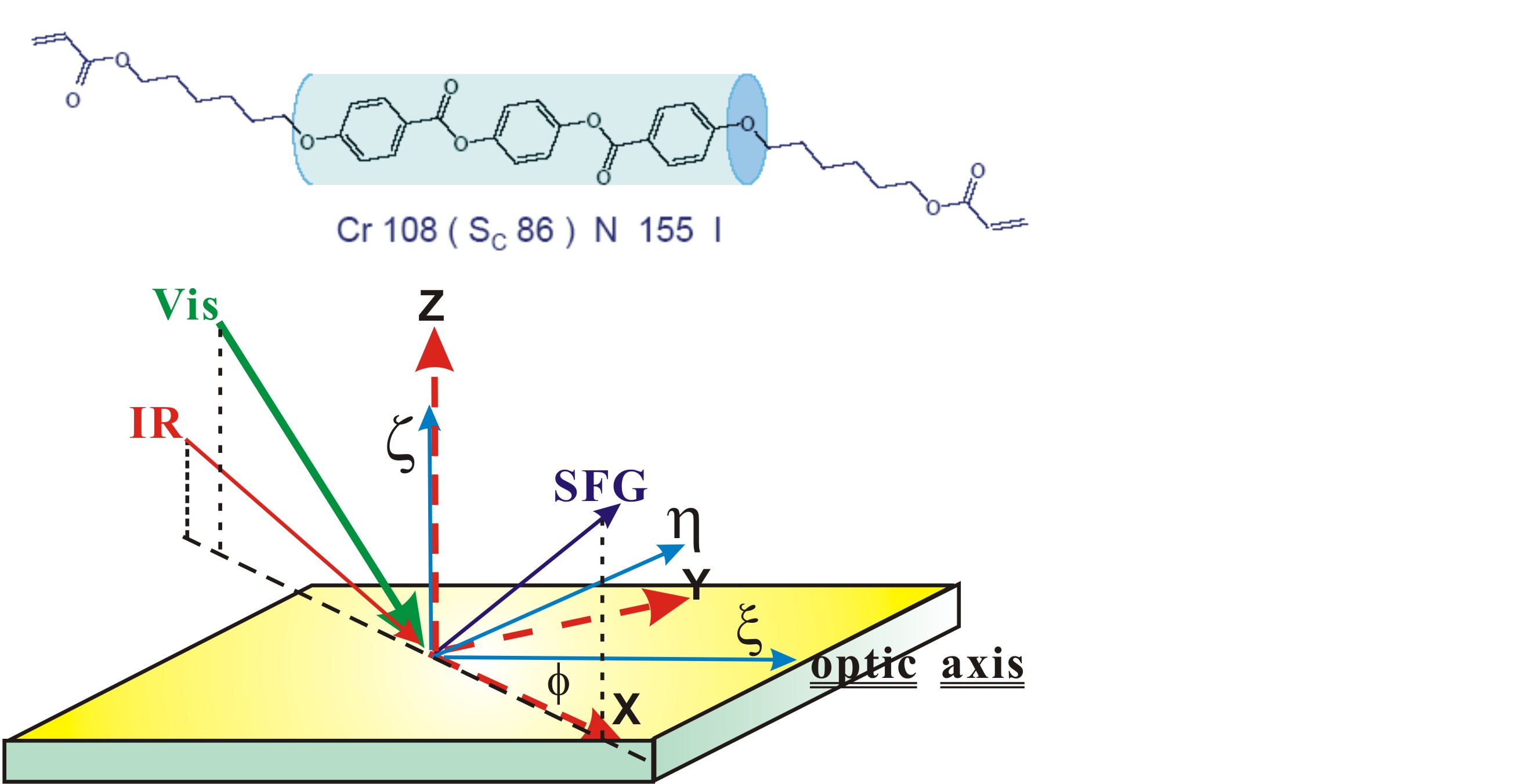
FIG. 8. Schematic angle-resolved SFVS depicting the relationship between the coordinate frames of the SFVS measurement (XYZ) and LPUV-irradiated layer. The inset above shows the molecular structure of the 1,4-phenylene bis(4-(6-(acryloyloxy) hexyloxy benzoate of liquid crystal polymer (LCP) moiety. The blue cylinder denotes the rod-like region of the LCP molecule. A. Polarization-resolved sum-frequency vibrational spectra (SFVS) of unrubbed SE7492 and LCP on unrubbed SE7492 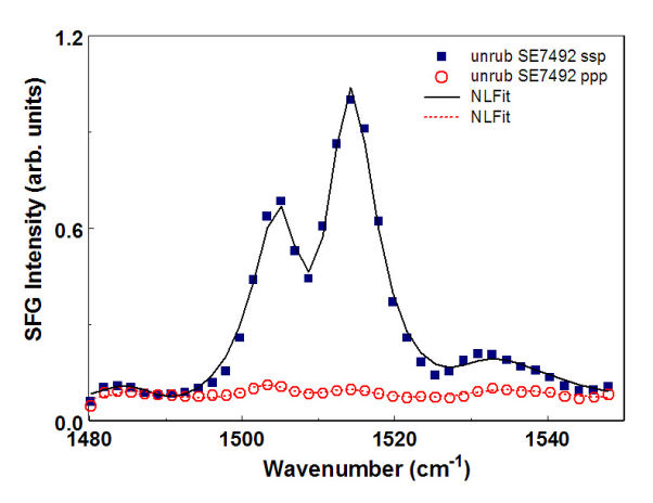 __
__
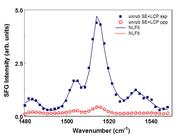
FIG. 9. ssp-Sum (a) Pol.-resolved SFVS of unrubbed SE7492 (Left). (b) Pol.-resolved SFVS of LCP on unrubbed SE7492 (Right). B. Angle-resolved ssp-SFVS of rubbed SE7492 and LCP on rubbed SE7492 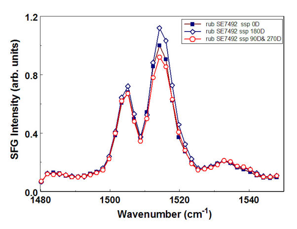 __
__
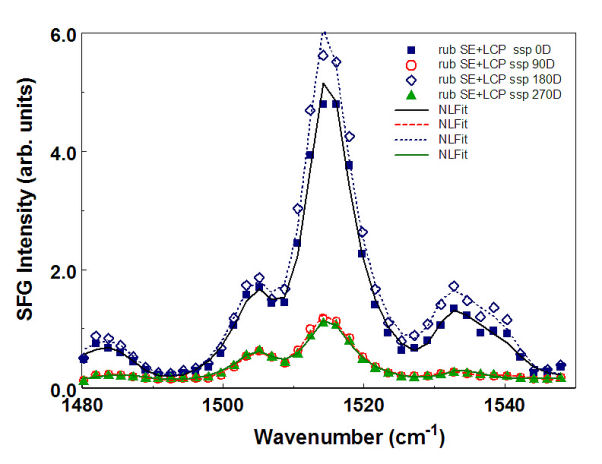
FIG. 10. (a) Angle-resolved ssp SFVS of rubbed SE7492. (b) Angle-resolved ssp SFVS of LCP on a rubbed SE7492. The polarization of the rubbing direction was oriented at 0 degrees , 90 degrees, 180 degrees, and 270 degrees relative to the incident plane of SFVS measurement. C. Polarization-resolved sum-frequency vibrational spectra of unexposed RN1349 and LCP on unexposed RN1349 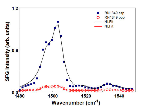 __
__
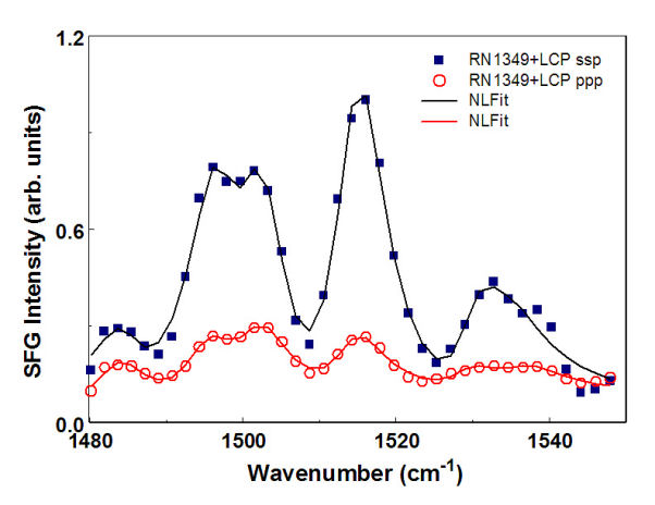
FIG. 11. (a) Pol.-resolved SFVS of unexposed RN1349. (b) Pol.-resolved SFVS of LCP on the unexposed RN1349. D. Angle-resolved ssp-sum-frequency vibrational spectra of UVA RN1349 and LCP on UVA RN1349 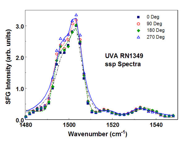 __
__

FIG. 12. (a) Angle-resolved ssp SFVS of LPUV-defined RN1349. (b) Angle-resolved ssp SFVS of LCP on the LPUV-defined RN1349. The electric field of LPUV light was oriented at 0 degrees, 90 degrees, 180 degrees, and 270 degrees relative to the incident plane of SFVS measurement. By employing sum-frequency vibrational spectroscopy, we demonstrated that the weak surface anisotropy of the phenyl ring in the LPUV-irradiated RN1349 layer was printed on a LCP overlayer and the resulting alignment order of the LCP phenyl rings were improved epitaxially with the layer thickness. |